Abstract
Approximately 60% of female, captive, white rhinoceroses (Ceratotherium s. simum) are acyclic.3 Recent ultrasound examinations, in combination with endocrine monitoring, have revealed ovarian pathologies such as cysts, micro-corpora lutea and inactive ovaries, all of which appear to be associated with lack of reproductive activity.1 In an attempt to treat these uterine and ovarian pathologies, long- and short-acting GnRH agonist implants (Deslorelin Implant®, Peptech Animal Health, Sydney, Australia; Ovuplant™ Deslorelin, Fort Dodge Animal Health, IA, USA) have been applied.2 As most rhinos examined in this study had not been trained to accept implant placement without anesthesia, we developed a remote delivery method using a dart originally developed by Telinject® (Veterinaer Spezialgeraete GmbH, Roemerberg, Germany) for the remote delivery of microchips.
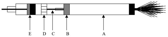
The dart consists of two distinct sections (Figures 1 and 2): A front delivery system with a pin that pushes the implant through the needle and an air chamber which on impact slides into the front part. The implant is placed in a needle normally used for microchip implantation. This needle is attached to the front of the dart. On impact, the pressurized 10 ml air in the air chamber is released as the valve (B) is impaled on pin (C). The air rushes through sieve (D) to push plunger and pin (E) forward into the needle. The pin pushes the implant out of the needle and it is deposited in the subcutaneous tissue.
Figure 1
On impact, air chamber (A) is pushed onto pin (C) opening rubber valve (B) and air rushes through sieve (D) to push plunger and pin (E) into needle and expulsing implant (see Figure 2).
Figure 2
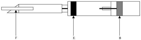
The air has pushed plunger (E) forward moving the pin into the needle and expulsing implant (F), depositing it in the subcutaneous tissue.
This dart system has been used successfully in numerous rhinoceroses to place deslorelin implants without the use of restraint or anesthesia. Long-term administration of GnRH agonist implants substantially improved the reproductive health status in aged female white rhinoceroses. This dart system constitutes an important additional tool in our remote delivery repertoire.
Literature Cited
1. Hermes, R., T.B. Hildebrandt, F. Schwarzenberger, C. Walzer, G. Fritsch, and F. Goeritz. 2001. Reproductive disorders in white rhinoceros and the value of ultrasonographic assessment. In: Schwammer, H.M., T.J. Foose, M. Fouraker, and D. Olson (eds.). A research update on elephants and rhinos. Proceedings of the International Elephant and Rhino Research Symposium, Schueling Verlag, Muenster, Germany. P. 314.
2. Hermes, R., C. Walzer, F. Schwarzenberger, F. Goeritz, M.L. Patton, K. Tomasova, and T.B. Hildebrandt. 2003. GnRH agonist implants for ovarian down regulation and ovulation induction in rhinoceroses. Verh. ber. Erkrg. Zootiere. 41:305–308.
3. Schwarzenberger, F., C. Walzer, K. Tomasova, J. Vahala, J. Meister, K.L. Goodrowe, J. Zima, G. Strauss, and M. Lynch. 1998. Faecal progesterone metabolite analysis for non-invasive monitoring of reproductive function in the white rhinoceros (Ceratotherium simum). Animal Reprod. Sci. 53:173–190.