July, 2001
This case is presented by Dr. Donald Levesque, Veterinary Neurological Center, Phoenix Arizona and Las Vegas Nevada;
with consultations by Dr. G. Diane Shelton and Dr. Alexander "Sandy" de Lahunta
Signalment, History, Physical Examination Neurological Examination Tests Performed Case Summary
Signalment, History, Physical Examination
 Signalment: Pomeranian, female, spayed; 4 years, 8 months old; habitat, Las Vegas, Nevada, U.S.A.
Past Medical Problems: None
History: History of one to one and one-half years of progressive weakness and paresis in pelvic limbs. Owner describes seizure-like episodes (generalized convulsions and focal seizures) and behavioral changes.
Physical Examination
General: Quiet, alert and responsive. Appears markedly paretic and has a crouching posture.
Body weight: = 4.1 kg. General body condition: Normal except for limb muscle atrophy.
Temperature: (rectal): 38.9C; Heart rate 100/min; Respiratory rate panting
Integument: N
EENT: N
Cardiopulmonary: Normal with synchronous pulses. Panting
Abdominal Cavity: N
Musculoskeletal: Moderate atrophy of limb musculature; no joint or long-bone pain.
Lymph Nodes: N
[Top]
Neurological Examination
State of Consciousness: Quiet, alert and responsive.
Gait and Posture: severe pelvic limb paresis, dragging rear limbs but with some movement possible; forelimbs appear to weaken with exercise.
Placing and Postural Reactions
Proprioceptive Placing Reactions (N=normal, S=slow, VS = very slow, A=Absent)
Thoracic limb N
Pelvic limb A
Hopping & Hemistand / Walk
Thoracic limb Left-N, Right-S (limb occasionally collapses)
Pelvic limb A
Wheelbarrow
Thoracic limbs - S
Pelvic limbs- A
Extensor Postural Thrust
Pelvic limb: Abnormal, limbs tend to drag, right limb more severely affected than left
Visual & Tactile Placing Reactions
Thoracic limb
Visual Left - Right -
Tactile Left - Right
Cranial Nerves
I: Not tested.
II: Normal visual fields. PLR - Equal pupils with intact direct and consensual responses. Normal menace reflex.
III, IV, VI: Normal physiological nystagmus present without positional nystagmus or strabismus. Pupils normal and of equal size. Pupillary light reflexes normal bilaterally.
V: Normal motor and sensory function present on both sides.
VII: Normal facial symmetry and normal movement of the muscles of facial expression.
VIII: Normal clinical response to auditory stimuli each ear. No head tilt, positional nystagmus or strabismus were noted.
IX, X, XI: Normal swallowing action was noted with stimulation.
XII: The tongue musculature was symmetrical and tongue movements were normal.
Spinal Reflexes: (Left/Right) (N=Normal, D=Decreased, I=Increased, A=Absent)
Thoracic Limb
Tendon Reflexes
Biceps N/N
Triceps N/N
Flexion Reflexes N/N
Crossed Extensor Reflexes N
Pelvic Limb
Tendon Reflexes
Patellar A/A
Flexion Reflexes N/N
Crossed-extensor Reflexes N/N
Perineal Reflexes N; anal tone normal
Cutaneous Trunci Reflexes N
Palpation of the head/neck/spine: Moderate atrophy of limb musculature. Normal range of motion. No hyperesthesia on palpation of spine.
[Top]
Tests Performed
Hematology, Chemistry and Urinalysis
Clinical Chemistry Laboratory Results
|
Constituent |
Patient's Results |
Units |
Reference Range (Dog) |
|
Alk. Phosphatase |
51 |
U/L |
23-212 U/L |
|
ALT (SGPT) |
28 |
U/L |
10-100 |
|
Ammonia |
|
mg/dl |
0-92 |
|
AST (SGOT) |
|
U/L |
15-43 |
|
Bile acids: |
|
fasting |
|
micromol/L |
0-12 |
|
post-prandial |
|
micromol/L |
0-16 |
|
Bilirubin: direct |
|
mg/dl |
0-0.1 |
|
Bilirubin: total |
0.17 |
mg/dl |
0-0.9 |
|
Blood urea nitrogen (BUN) |
18.1 |
mg/dl |
7.0-27.0 |
|
BUN/creatinine ratio |
|
|
6-25 |
|
Calcium |
10.17 |
mg/dl |
7.90-12.0 |
|
Cholesterol |
159.4 |
mg/dl |
110-320 |
|
Creatine kinase |
|
|
|
|
Clotting: |
|
PT |
|
SEC |
7.5-19.5 |
|
PTT |
|
SEC |
9-12 |
|
PIVKA |
|
SEC |
15-18 |
|
FDP |
|
|
<10 |
|
Creatine |
0.58 |
mg/dl |
0.5-1.80 |
|
Creatine kinase |
|
U/L |
46-320 |
|
Creatinine |
|
mg/dl |
0.8-1.6 |
|
Glucose |
123 |
mg/dl |
77-125 |
|
Electrolytes: |
|
Anion gap |
|
mmol/l |
12-25 |
|
Chloride |
114 |
mmol/l |
109-122 |
|
CO2, total |
|
mmol/l |
16-26 |
|
Potassium |
4.66 |
mmol/l |
3.50-5.80 |
|
Sodium |
156 |
mmol/l |
144-160 |
|
Lipase |
|
U/L |
0-500 |
|
Magnesium |
|
mg/dl |
1.2-2.4 |
|
Phosphorus, inorganic |
3.20 |
mg/dl |
2.5-6.80 |
|
Proteins: |
|
Albumin |
3.39 |
g/dl |
2.70-3.80 |
|
A/G ratio |
|
|
0.6-1.2 |
|
Globulin |
3.53 |
g/dl |
2.50-4.50 |
|
Total protein |
6.92 |
g/dl |
5.20-8.20 |
|
Thyroid: |
|
Thyroxine T4 |
|
micrograms/dl |
1.0-3.6 |
|
Free T4-EQ.D. |
|
ng/ml |
1.0-3.5 |
|
TSH-Canine |
|
mU/L |
2-30 |
|
Tri-iodothyro. T3 |
|
ng/dl |
75-150 |
|
Triglycerides |
|
mg/dl |
19-133 |
Hemogram Results
|
|
Patient's results |
Reference Values (Dog) |
|
Erythrocytes |
6.93 |
5.5-8.5 million |
|
Hemoglobin (Hb) |
19.6 |
12.0-18.0 g/dl |
|
Hematocrit |
55 |
37-55% |
|
Mean corpuscular volume |
69.3 |
62-77 fl |
|
Mean corpusc. Hb |
24.8 |
33-37 g/d |
|
Mean corpusc. Hb conc. |
35.6 |
21.5-26.5 pg |
|
Reticulocytes |
0.3 % |
0.5-1 % |
|
Leucocytes |
10400 |
6000-17000/microliter |
|
Band |
|
0-300/microliter |
|
Neutrophils |
8000 (77%) |
3000-11500/microliter |
|
Lymphocytes + Monocytes |
2400 |
1000-4800/microliter |
|
%L/M |
23 |
|
|
Monocytes |
|
150-1350/microliter |
|
Eosinophils |
5500 |
100-1250/microliter |
|
Basophils |
0 |
Rare |
|
Platelets |
215,000 |
200-500x1000 |
|
Icteric Index |
|
2.0-5.0 |
|
Plasma proteins |
7.4 |
6.0-8.0 |
|
Fibrinogen |
300 |
200-400 mg/dl |
|
Protein:fibrinogen |
|
>15:1 |
Acetylcholine receptor antibody - Titer: Negative
Cerebrospinal Fluid: Total and Differential Cell Counts; Total Protein
Fluid from: Cerebello-medullary cistern
Gross appearance: Clear
Refractive index: 1.3350
Total Protein: 118 mg/liter
Total erythrocytes:
Total nucleated cells: 8/microliter
Differential Nucleated Cell Counts
Smear type:
Neutrophils: 0%
Small mononuclear cells:
Large mononuclear cells:
Eosinophils: 0%
Microscopic evaluation: Lymphocytes, occasional clumps of macrophages
Clinical Neurophysiological Examinations
Electromyography Results
Bipolar, coaxial needle electrodes were used to record the EMG of the head, limbs and paraspinal muscles while the dog was anesthetized with thiopental. Fibrillation potentials and positive sharp waves were found in all limb muscles but were most pronounced in the pelvic limbs.
Motor Nerve Conduction Velocity
Ulnar Nerve: left = 13.2 m/s; right = 40.4 m/s
Sciatic nerve: left = 16/3 m/s (Fig A); right = 40.6 m/s
Click on the image to see a larger view.
| Figure A. | 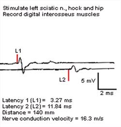
|
|
| |
|
Figure A. Nerve conduction velocity recording. Note small action potentials with extremely slow conduction velocity and dispersion. |
Biopsy
Muscle and Nerve Biopsy Report
G. Diane Shelton
Stains Employed. For muscle specimens: Hematoxylin and eosin, modified Gomori's trichrome, periodic acid Schiff's, adenosine triphosphatases at pH 9.8 and 4.3, non-specific esterase, nicotinamide adenine dinucleotide-tetrazolium reductase (NADH-TR), acid phosphatase, alkaline phosphatase, oil red 0, staphylococcal protein A-horseradish peroxidase (SPA-HRPO). For nerve specimens: Toluidine blue.
Fresh and fixed biopsies were submitted from the quadriceps and extensor carpi radialis muscles. There was generalized myofiber atrophy within the quadriceps muscle with an extensive type 1 fiber predominance. Intramuscular nerve branches were moderately depleted of myelinated nerve fibers. Excessive lipid droplets were present within type 1 fibers. A moderate variation in myofiber size was present within the ECR with scattered singular angular atrophied fibers of both fiber types and large groups of atrophic fibers of both fiber types.
Fresh and fixed biopsies were submitted from the left ulnar and sciatic nerves for evaluation. The fixed biopsies were plastic embedded and evaluated in 1 micrometer sections. Mild axonal degeneration with occasional myelin ovoids were present within the ulnar nerve. More marked axonal degeneration and numerous myelin ovoids were present within the sciatic nerve. Non-suppurative cellular infiltration, most marked within the sciatic nerve, was present in a sub-perineurial and endoneurial distribution. Regeneration was not observed.
Conclusion: Axonal degeneration and non-suppurative cellular infiltration most marked within the sciatic nerve and present to a much lesser extent within the ulnar nerve. While the type 1 fiber predominance within the quadriceps muscle is suggestive of significant reinnervation, nerve fiber regeneration was not prominent. The axonal degeneration and cellular infiltration are likely an extension of the polyradiculoneuritis.
Click on an image to see a larger view.
|
|
Extensor carpi radialis muscle.
There are scattered angular and anguloid fibers of both fiber types (ATPase not shown) consistent with denervation. |
|
|
Ulnar nerve, plastic embedded nerve biopsy section, toluidine blue stain.
No cellular infiltration obvious, with approximately normal density of myelinated fibers. |
|
|
Sciatic nerve, plastic embedded nerve biopsy section, toluidine blue stain.
Non-suppurative cellular infiltration in a subperineurial distribution (arrows) and scattered throughout the endoneurium. Nerve fiber loss, axonal degeneration and myelin ovoids are present also. |
|
|
Sciatic nerve, plastic embedded nerve biopsy section, toluidine blue stain.
Cluster of cellular infiltrates within the endoneurium. Also obvious is nerve fiber loss and myelin ovoids. |
[Top]
Case Summary
Additional Diagnostic Investigations, and Final Diagnosis
Summary: A four-year-old ovariohysterectomized female Pomeranian was examined for evaluation of a 12 to 18 month history of chronic progressive pelvic limb paresis and weakness. The owners reported the onset of seizure-like activity and behavioral changes within the last 2 months. On physical examination, the dog was weak in all four limbs, worse in the pelvic limbs. Her rear limbs tended to drag although she had voluntary motor activity and made walking motions. Conscious proprioception was absent in the rear limbs, as were both patellar and withdrawal reflexes. The withdrawal reflexes were also weak in the front limbs, especially on the right. Mentation and cranial nerve examination was normal. General physical examination was unremarkable. The primary rule outs included diffuse or multifocal myelopathy or polyneuropathy. The owners opted in favor of euthanasia due to the expense and overall poor prognosis. They granted permission for electrophysiologic testing, cerebrospinal fluid collection and nerve and muscle biopsies prior to euthanasia and necropsy.
The results of all tests are presented in the various pages above.
The clinical signs, EMG, nerve conduction velocity tests and CSF examinations all yielded results consistent with a polyneuropathy, The nerve and muscle biopsies and microscopic examinations of the brain and spinal cord confirmed this diagnosis and provided additional important information about the nature of the polyneuropathy that afflicted this unfortunate dog.
Dr. Shelton reported that the nerve biopsies showed non-suppurative cellular infiltrations, nerve fiber loss, axonal degeneration and myelin ovoids in both the sciatic and ulnar nerves, more pronounced in the sciatic nerve.
Dr. de Lahunta reported similar findings in nerve roots of the spinal cord and brain and provided additional information about the nature of the disease. His report follows.
Report on gross and microscopic examinations of the brain and spinal cord.
Dr. Alexander (Sandy) de Lahunta
There were no gross lesions on the external surface of the brain or spinal cord. On the transverse sections of the cervical spinal cord there was a very slight white discoloration in the dorsal funiculi. On microscopic examination there is a very severe extensive diffuse non-suppurative radiculitis of both dorsal and ventral roots and all components of the spinal nerves that I sectioned including some involvement of the spinal ganglia (Figure A). The destruction of the myelin and axons is profound although the former is easier to appreciate.
Click on the image to see a larger view.
|
|
Figure A. Dorsal root ganglion/dorsal root. Hematoxylin and eosin stained.
Severe non-suppurative cellular infiltration. |
The only lesion in the spinal cord is a mild degeneration in the dorsal funiculi from the loss of dorsal root axons that are proprioceptive and pass into the dorsal funiculi to ascend to the caudal medulla before synapsing. This is just wallerian degeneration of these central processes and is the basis for the white discoloration that I saw on gross examination. This part of the lesion confirms that there is considerable axonal loss from the radiculitis. Although most all of the roots are affected the lesion is most extensive in the lumbosacral area.
On the surface of the brain where the cranial nerves attach there is a similar radiculitis especially of the oculomotor neurons but there are mild lesions of most of the ones that were present. The wallerian degeneration in the spinal tract of V in the medulla indicates that there are lesions in the trigeminal sensory neurons.
I looked long and hard for protozoal organisms and found none but have requested immunocytochemistry just to be sure.
In the absence of any infectious agent, I interpret these lesions as a chronic immune-mediated polyradiculoneuritis. The fact that the lesion stops at the CNS - PNS boundary (Figure B) exactly where the central oligodendroglial myelin changes to the Schwann cell myelin of the peripheral nerves supports this as an immune-mediated disorder primarily directed against Schwann cell myelin and secondarily the axons. If this radiculitis were caused by Neospora there would be at least some involvement of the spinal cord parenchyma.
Click on the image to see a larger view
|
|
Figure B. Spinal cord and radicular rootlets. Hematoxylin and eosin stained.
Cellular infiltration of peripheral rootlets (e. g., blue arrows) stops at the peripheral nervous system/central nervous system boundary (red arrow) |
I could find no brain lesion to associate with the seizure-like activity that was reported for this dog.
Final diagnosis: poly-radiculopathy/polyneuropathy, inflammatory, probably immune mediated. This case demonstrates emphatically the severity such neuropathies can reach and suggests their ultimate end point if indeed they are allowed to go that far.
[Top]