Bernd Tellhelm, Dr.med.vet. DECVDI
Normal Elbow Joint
(from Waibl et al.: Atlas of Radiographic Anatomy of the Dog, Parey 2003)
A Humerus
B Radius
C Ulna
2 medial humeral condyle
4 lateral epicondyle
6 medial epicondyle
13 medial coronoid process
14 lateral coronoid process
16 anconeal process
|
| Mediolateral view | 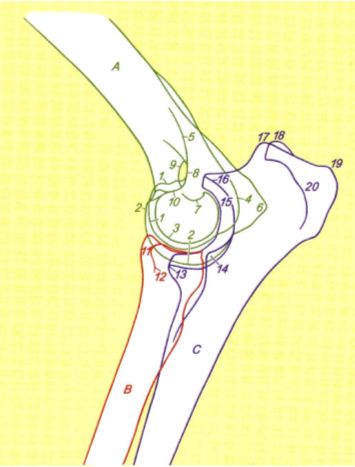
|
|
| |
|
3 medial humeral condyle
7 lateral coronoid process
8 medial coronoid process
|
| Craniocaudal view | 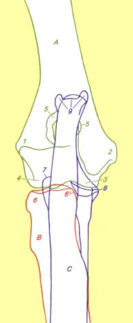
|
|
| |
|
Developmental phases of the canine Elbow
Ossification centers of
1. humeral condyle
2. medial epicondyle (anconeal process not yet visible!)
3. proximal radial epiphysis
Primary ED-Lesions (IEWG)
 Ununited Anconeal process (UAP)
Fragmented medial coronoid process (FCP)
Osteochondritis (dissecans) medial humeral condyle (OCD)
Severe Incongruity/step between radius and Ulna (Inc)
For radiographic details please refer to the article by Flückiger. "Radiographic Diagnosis of Elbow Dysplasia in the Dog"