Laboratory of Veterinary Pathology, Graduate School of Agricultural and Life Sciences, The University of Tokyo
Tokyo, Japan
Senile plaques (SP) and amyloid angiopathy seen in the Alzheimer's disease (AD) brain have been found in the brains of aged dogs (more than 9 years old) (Figs. 1 and 2). However, neurofibrillary tangles (NFT), another characteristic lesion in AD, have never been observed in the brains of aged dogs. Neuronal cell loss is an additional histopathological hallmark and apoptotic neuronal cell death was detected in the brains of AD patients. It would be worthwhile to clarify the difference of brain lesions between AD patients and other aged mammalian species including dogs and to discuss the usefulness of aged mammals as models for AD. Therefore, we examined brain pathology in the aged animal brains.
Apoptotic (TUNEL stain-positive) cells were present in both the cortex and white matter of the aged dog brain (Fig.3). Apoptotic neurons were swollen and larger than intact cells, although these changes were slight. Apoptotic bodies or chromatin margination, which are the typical morphological characteristics of apoptosis, were not observed. TUNEL-positive cells in the brain were revealed to be astroglia and oligodendroglia as well as neurons (Fig.3). These observations suggest that a wide variety of brain cells participate in the pathological changes in aged dogs.
The number of TUNEL-positive cells and the number of SP tended to increase with age (Fig.4 a and b). On the contrary, the numbers of SP and TUNEL-positive cells showed no correlation (Fig.4c). The examination of aged dogs clinically evaluated for dementia before death using the index (dementia index, DI) system, revealed a significant positive correlation between the number of TUNEL-positive cells and the severity of dementia (Fig.5a). However, there was no relationship between the number of SP and DI (Fig.5b). The results suggest that the two age-related lesions (apoptosis and SP) could independently occur. Furthermore, brain cell apoptosis rather than SP might be more appropriate histopathological hallmark accounting for canine dementia.
Apoptotic lesions in the frontal brain were the most crucial for the dementia condition. The histochemistry for detecting antioxidant enzymes revealed that the number of superoxide dismutase (SOD)-depleted neurons and SOD-positive glial cells increased in an age-dependent manner. The free radical-mediated cell injury might play a crucial role(s) for brain cell apoptosis in aged dogs.
Above mentioned aged-related brain pathology was detected not only in mammalian but also avian species (Fig.6). A variety of SP morphology was observed according to species (Figs.6 and 7). SP of dogs and monkeys were divided into 2 types, diffuse (DP) and mature (MP) types as in human. The shape of SP of the cat and camel are very different from that of other species. Amyloid angiopathy was detected also in several species. However, NFT which had been reported only in the sheep, bear, cattle and wolverine, was never observed in the species examined in the present study.
The aged mammalian species, in particular dogs, would be useful animal models to elucidate the pathomechanism of Alzheimer's disease.
Click on image to see a larger view
| 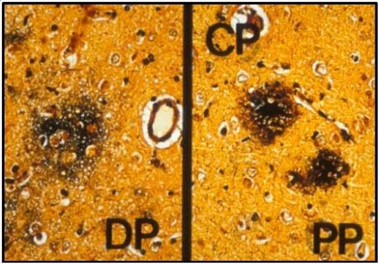
Fig. 1 Senile plaques of the dog. Diffuse (left) and mature (right) plaques. PAM stain. |
|
| |
Click on image to see a larger view
| 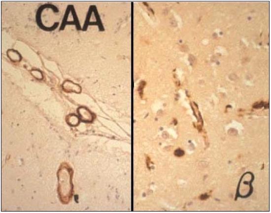
Fig. 2 Amyloid angiopathy (amyloid deposition in the vessel wall) in the brain of the aged dog. Immunohistochemistry for beta amyloid protein. |
|
| |
| 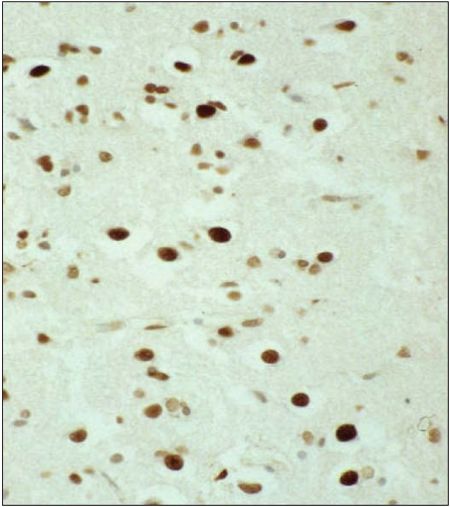
Fig. 3 Apoptotic brain cells (TUNEL-positive cells) in an aged dog brain. |
|
| |
Click on image to see a larger view
| 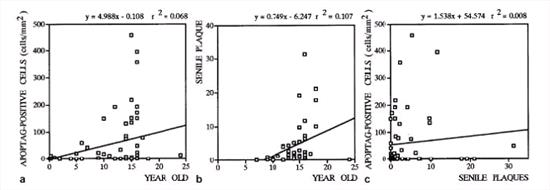
Fig. 4 Apoptosis in the aged dog brain. Correlation between apoptotic cell number and age (a), that between SP number and age (b) and that between apoptotic cell number and SP number (c). |
|
| |
Click on image to see a larger view
| 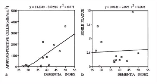
Fig. 5 Correlation between the number of apoptotic (APOPTAG (TUNEL)-positive) cells and dementia index (a) and correlation between the number of senile plaques and dementia index (b). |
|
| |
Click on image to see a larger view
| 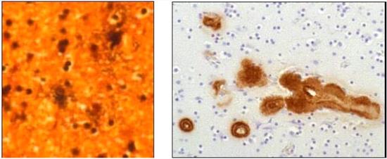
Fig. 6 Senile plaque-like structure (left, PAM stain) and amyloid angiopathy (right, immunohistochemistry for beta amyloid) in the brain of a woodpecker. |
|
| |
Click on image to see a larger view
| 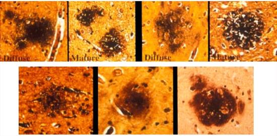
Fig. 7 Senile plaques of various mammalian species. Upper, left to right: Dog (DP), dog (MP), monkey (DP) and monkey (MP). Lower, left to right: Cat, camel and bear. PAM stain. |
|
| |