James W. Buchanan, DVM, M Med Sci, DACVIM
This communication presents angiocardiograms and related images of congenital cardiovascular abnormalities. Many of these were published in the references below. Viewers who want to look at specific abnormalities without self-testing can click here to see a list of the figures in this site. Acquired abnormalities are shown separately in Angiocardiography 101-acquired. Additional images of some congenital abnormalities will be presented in separate sites under development. Clinical features and surgery for some conditions are presented in the Small Animal Cardiac Surgery collection.
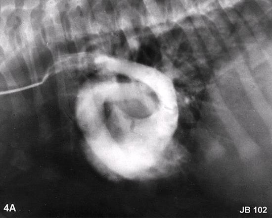
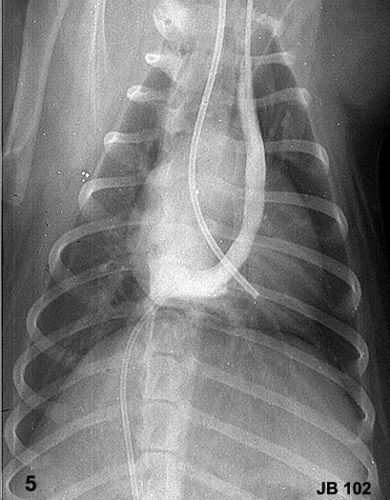
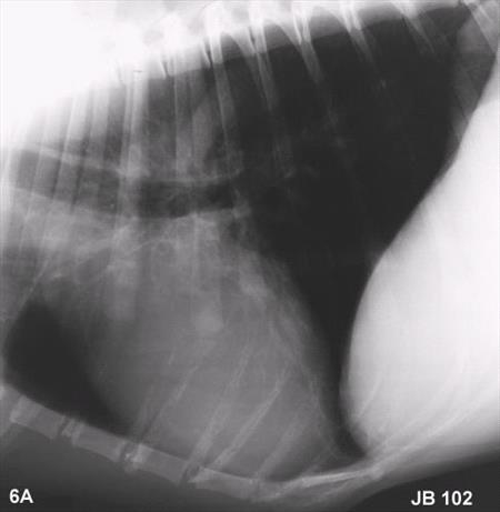
For self-study and testing, all angiographic images are presented without labels then duplicated with labels and captions. Readers are encouraged to determine where the contrast material (Cardiografin 85% or Renografin 76%, 0.5cc/lb, or @1cc/kg)] was injected and what abnormality was demonstrated before advancing to the labeled image and caption.
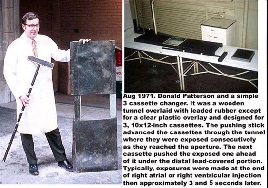
Figure one is a diagram of the circulation, normal pressures and common shunts. Self-test viewers should print the page and try to add labels to all parts of the circulation along with normal systolic and diastolic pressures. The pressures determine the direction and timing of blood flow across shunts and valves and guide optimal catheter positioning for selective angiocardiography. Figure two shows an inexpensive cassette changing device that was used to make several of the angiocardiograms in this series before more sophisticated and expensive equipment became available (1).
Former residents and colleagues at the University of Pennsylvania are acknowledged for performing some of the angiographic studies in this site.
References:
- Patterson DF, and Botts RP. A simple cassette changer. Sm An Clin. 1:1-11, 1961
- Buchanan JW, Detweiler DK, and Hubben K:Clinico-Pathologic Conference: Pulmonic stenosis and patent foramen ovale in a dog. J Am Vet Med Assoc 139:701-707, 1961.
- Buchanan JW: Persistent left cranial vena cava in dogs: Angiocardiography, significance and co-existing anomalies. J Am Vet Radiol Soc (now Vet Radiol and Ultrasound) 4:1-8, 1963.
- Buchanan JW: Selective Angiography and Angiocardiography in Dogs with Acquired Cardiovascular Disease. J Am Vet Radiol Soc 6:5-20, 1965.
- Buchanan JW, and Patterson DF: Selective angiography and angiocardiography in dogs with congenital cardiovascular disease. J Am Vet Radiol Soc 6:21-39, 1965.
- Buchanan JW, and Pyle RL: Cardiac tamponade during catheterization of a dog with congenital heart disease. J Am Vet Med Assoc 149:1056-1066, 1966.
- Buchanan JW: Patent ductus arteriosus and persistent right aortic arch surgery in dogs. J Sm An Pract 9:409-428, 1968.
- Lombard CW, Knight DH, Buchanan JW, and Riffle RA: Aortopulmonary window in a dog. A clinicopathologic conference. J Am Vet Med Assoc 172:75-80, 1978.
- Buchanan JW: Pulmonic stenosis caused by single coronary artery in dogs: 4 cases (1965-1984). J Am Vet Med Assoc196:115-120, 1990.
- Sammarco CD, Regan J, Ward CR and Buchanan JW: Caudal venous return through a left azygous vein in a dog. Vet Radiol. and Ultrasound 36:517-522, 1995.
- Buchanan JW. Patent ductus arteriosus: Morphology, pathogenesis, types and treatment. Jr Vet Cardiology 3;7-16, 2001.
- Buchanan JW. Tracheal signs and associated vascular anomalies in dogs with persistent right aortic arch. Jr Vet Intern Med. 18:510-514, 2004
Figure 1

Click
here to see the answer.
Figure 2
Click
here for example.
Click
here to see the answer.
Figure 3

Click
here to see the answer.
Figure 4
Click
here to see the answer.
Figure 5
Click
here to see the answer.
Click
here for flow patterns.
Click
here for surgical images.
Figure 6
Click
here to see vein injection.
Click
here to see the answer.
Figure 7
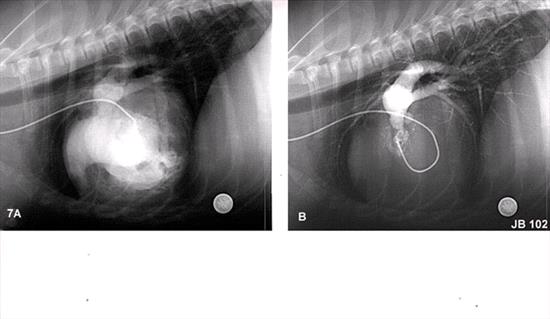
Click
here to see the answer.
Click
here for necropsy image.
Figure 8
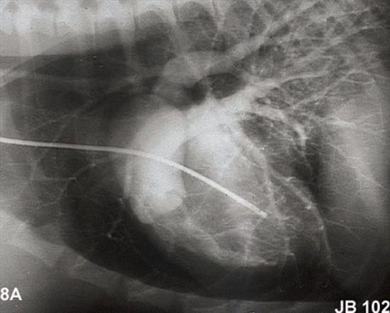
Click
here to see the answer.
Click
here for diagram.
Click
here for necropsy image.
Click
here for DV image.
Click
here for balloon image.
Figure 9
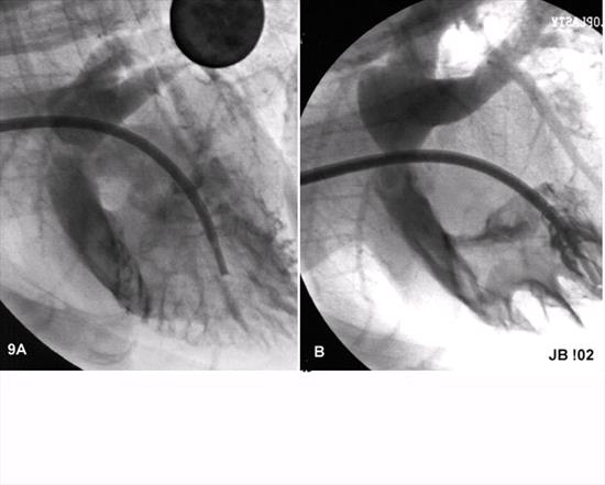
Click
here to see the answer.
Figure 10
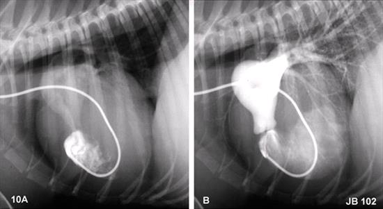
Click
here to see the answer.
Figure 11
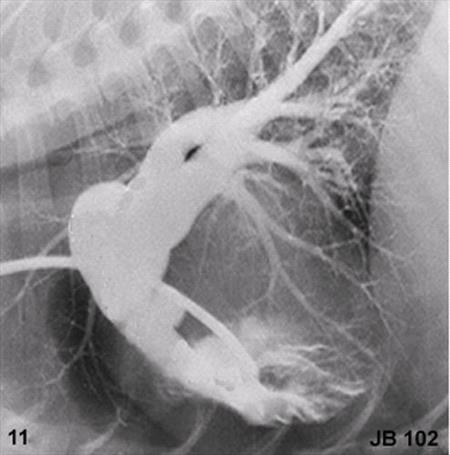
Click
here to see the answer.
Figure 12
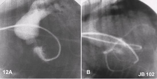
Click
here to see the answer.
Click
here for diagram.
Click
here for necropsy image.
Figure 13
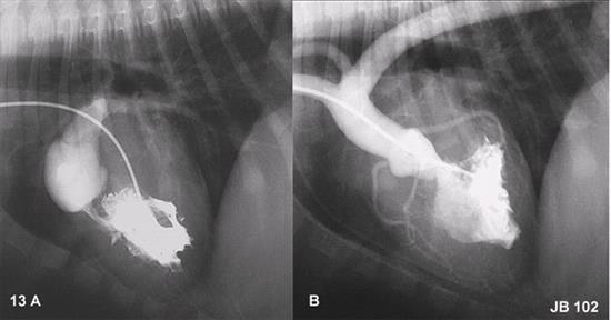
Click
here to see the answer.
Click
here for necropsy images.
Fgiure 14
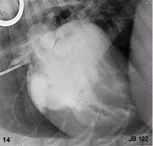
Click
here to see the answer.
Figure 15
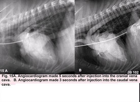
Click
here to see the answer.
Click
here for necropsy images.
Click
here for necropsy image.
Figure 16
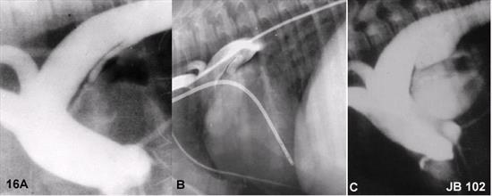
Click
here to see the answer.
Click
here for necropsy images.
Click
here for coil images.
Figure 17
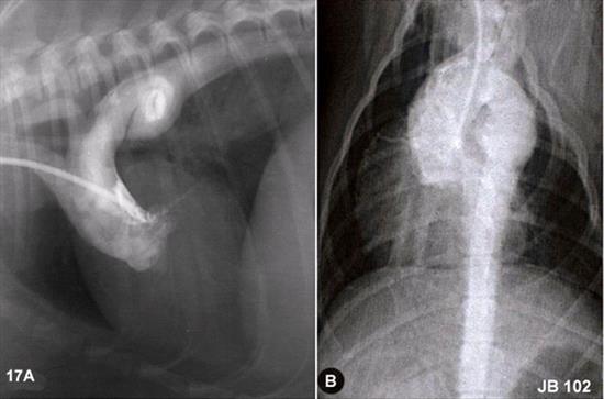
Click
here to see the answer.
Click
here for surg image.
Click
here for alternate image.
Figure 18
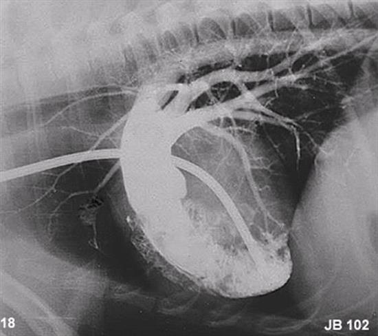
Click
here to see the answer.
Click
here for histology.
Click
here for lung study.
Figure 19
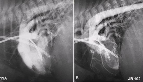
Click
here to see the answer.
Figure 20
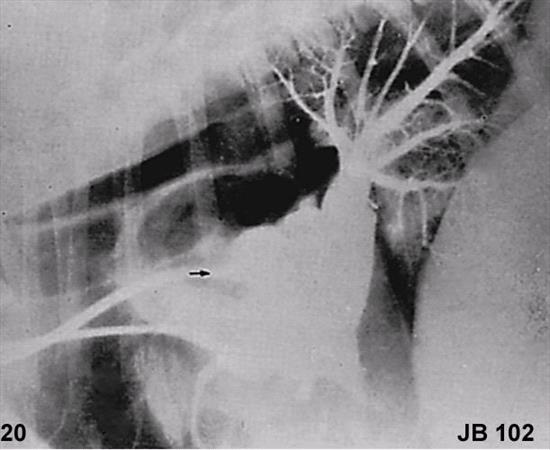
Click
here to see the answer.
Figure 21
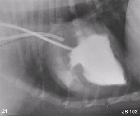
Click
here to see the answer.
Figure 22
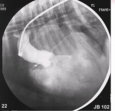
Click
here to see the answer.
Click
here for necropsy image.
Figure 23
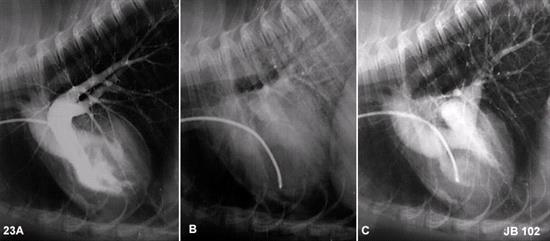
Click
here to see the answer.
Figure 24
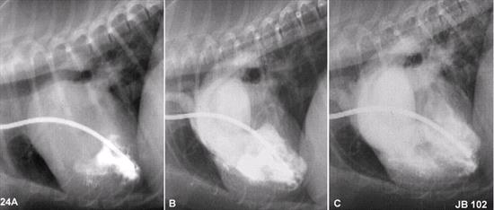
Click
here to see the answer.
Click
here for necropsy image.
Figure 25
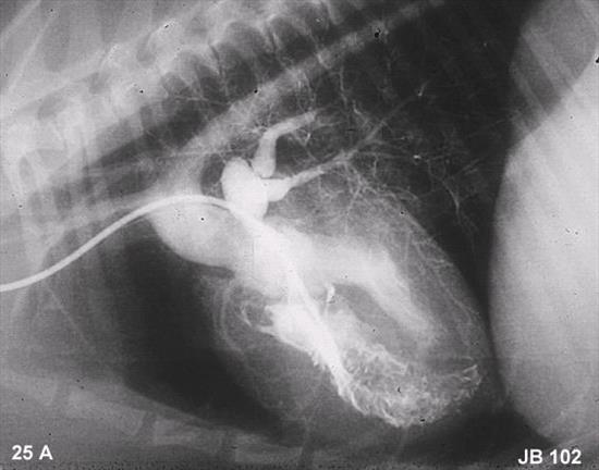
Click
here to see the answer.
Click
here for necropsy image.
Figure 26
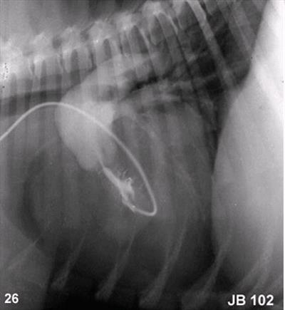
Click
here to see the answer.
Figure 27
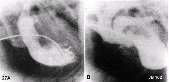
Click
here to see the answer.
Click
here for diagram.
Figure 28
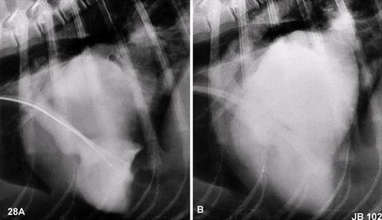
Click
here to see the answer.
Figure 29
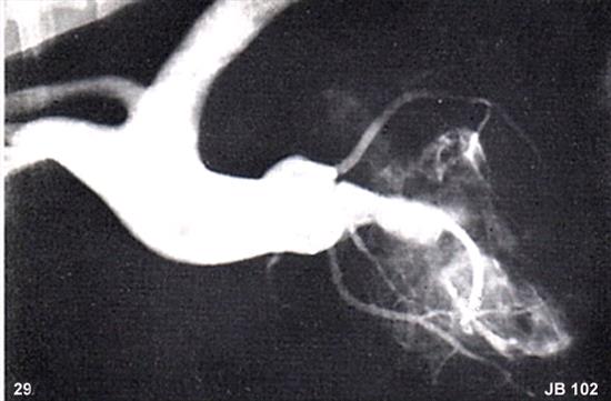
Click
here to see the answer.
Figure 30
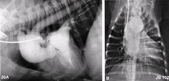
Click
here to see the answer.
Click
here for necropsy image.
Figure 31
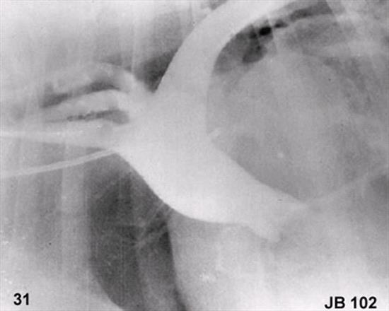
Click
here to see the answer.
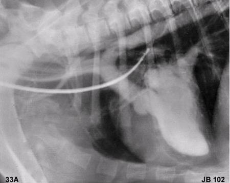
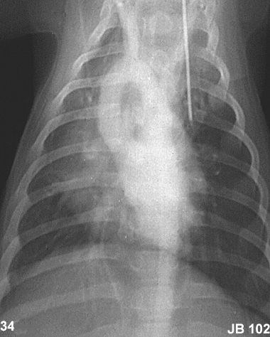
Figure 32
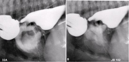
Click
here to see the answer.
Click
here for radiograph.
Click
here for diagram.
Click
here for tracheal radiographs.
Click
here for histology.
Click
here for co-existing anomalies.
Figure 33
Click
here to see the answer.
Click
here for diagram.
Figure 34
Click
here to see the answer.
Figure 35
Click
here to see the answer.
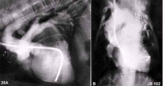
Figure 36
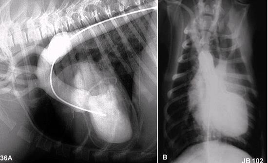
Click
here to see the answer.
Click
here for surgery image.
Click
here for surgery images.
Click
here for radiology images.
Click
here for patient image.
Figure 37
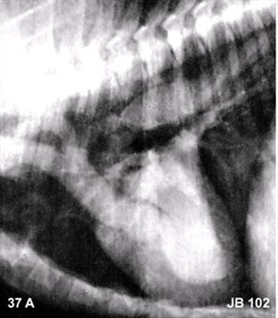
Click
here to see the answer.
Click
here for necropsy image.
Figure 38
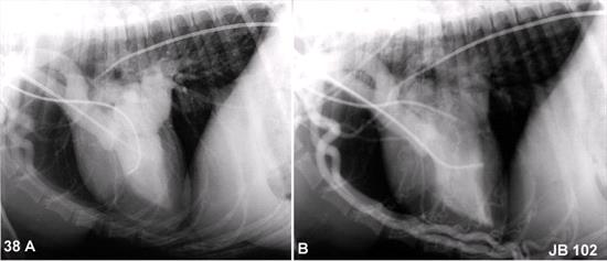
Click
here to see the answer.
Click
here for aortogram.
Click
here for necropsy image.
Click
here for necropsy images.
Click
here for diagram.
Figure 39
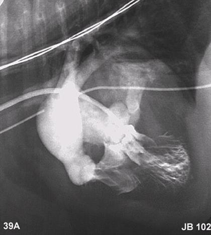
Click
here to see the answer.
Click
here for additional angios.
Click
here for necropsy images.
Click
here for necropsy image.
Figure 40
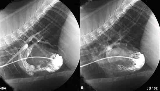
Click
here to see the answer.
Click
here for necropsy image.
Click
here for necropsy image.
102 Angiocardiography - Congenital - list of figures
Click on the name to see the figure.
- Circulation diagram
- Simple cassette changer
- Normal angiocardiogram
- Persistent left cranial vena cava
- Two venae cavae
- Left azygos vein
- Tricuspid dysplasia
- Pulmonic stenosis-valvular
- Pulmonic stenosis-supra- and subvalvular
- Pulmonic stenosis-immediate subvalvular
- Pulmonic stenosis-valvular and infundibular
- Pulmonic stenosis-single right coronary artery
- Double chambered right ventricle
- Pulmonic stenosis and atrial septal defect
- Pulmonic stenosis and patent foramen ovale
- Patent ductus arteriosus
- Ductal aortic aneurysm
- Patent ductus arteriosus with right-to-left shunt
- Patent ductus arteriosus bidirectional
- Atrial septal defect
- Ventricular septal defect
- Ventricular septal defect and aortic insufficiency
- Ventricular septal defect and pulmonic stenosis
- Double outlet right ventricle
- Tetralogy of Fallot
- Pulmonic stenosis-infundibular
- Aorticopulmonary window
- Mitral regurgitation
- Subaortic stenosis-mild
- Subaortic stenosis-severe
- Hypoplastic aortic root
- Persistent right aortic arch
- Retroesophageal left subclavian artery
- Retroesophageal left subclavian artery-DV
- Persistent right aortic arch and PDA
- Double aortic arch
- Coarctation of the aorta
- Interrupted aorta
- Cor triatriatum dextra
- Cor triatriatum sinister