Diagnosis of Malignant Nasopharyngeal Histiocytoma in a Domestic Shorthair Cat
Malignant histiocytic tumors are rarely seen in cats. To the author's knowledge, this is the first report of malignant nasopharyngeal histiocytoma in a cat. A 12 years and 10 months old female spayed domestic short hair cat was presented with a history of coughing, retching and loss of appetite. Apart from mildly reduced skin turgor, the clinical examination was unremarkable. Blood tests revealed elevated total protein, all other parameters were within normal limits. X-rays of the skull were taken and showed increased opacity and widening of the nasopharynx (Figure 1). Retrograde rhinoscopy was performed and a nasopharyngeal tumor located at the level of the soft palate was discovered (Figure 2). Multiple biopsies were obtained. For the histologic examination the biopsies were fixed in formalin, embedded in paraffin wax, sectioned and stained with H & E. Besides inflammatory infiltration a neoplastic cell population resembling atypic histiocytes could be demonstrated (Figure 3). Neoplastic cells stained negative for Cytoceratine-Ag, S-100-Ag and Toluidine-Blue. Positive Vimentin- and HLA-immunostaining confirmed the mesenchymal and histiocytic nature of the neoplasm (Figures 4 and 5). The application of immunohistochemical methods assisted the diagnosis of malignant histiocytoma in this case as routine H & E - staining did not yield distinct results.
Conclusions
 As nasal neoplasia is a common finding in cats, importance has to be placed on accurate and early diagnosis in order to balance prognosis and therapy.
Tentative diagnosis of nasal neoplasia demands the application of imaging techniques.
Endoscopy features the advantages of a low stress procedure combined with high visibility and the possibility to obtain biopsy samples.
Immunohistochemical stainings can be very useful in identification of poorly differentiated, high-grade malignant and uncommon tumors.
| Figure 1. | 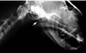
|
|
| |
| Figure 2. | 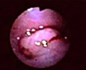
|
|
| |
| | Figure 3. | 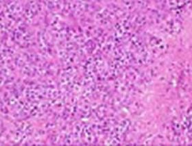
|
|
| |
|
| Figure 4. | 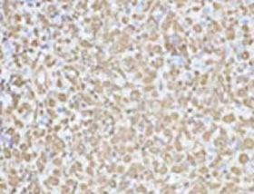
|
|
| |
| | Figure 5. | 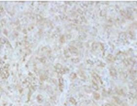
|
|
| |
|