July, 2001
Signalment, History, Physical Examination Neurological Examination Tests Performed Case Summary
Signalment, History, Physical Examination
 Signalment: This patient is a three-year-old, spayed female, domestic short-haired cat with a five month history of progressive pelvic limb weakness and reluctance to jump. Previously the patient had similar clinical signs which responded to a steroid injection.
Past Medical History: Previous history of popliteal lymphadenopathy, fine needle aspirates were suggestive of reactive lymph nodes. No other history of previous trauma or illness was reported.
Medications: No medications are being given at this time
Physical Examination:
General: Weight 7.58 lbs, Temperature 103, Pulse 140/BPM, Regular rhythm with pulses equal & synchronous, Respiratory rate 40/min, eupneic and mucous membranes pink, Capillary refill time < 2 sec. Alert & Responsive, Body Condition Score 5.5 / 9
Musculoskeletal: Mild to moderate discomfort with flexion & extension of both hocks, stifles and carpi. Mild effusion both tibiotarsal joints.
Lymph Nodes: Both Popliteal lymph nodes were 2.5- 3 cm non-painful to palpation
[Top]
Neurological Examination
State of Consciousness: Alert & Responsive
Gait: The cat's gait could not be fully examined but it was believed there was at least a suggestion of generalized weakness in all limbs with the pelvic limbs somewhat more affected then the thoracic limbs.
See video

Placing and Postural Reactions
Proprioceptive Placing Reactions (N=normal, S=slow, VS = very slow, A=Absent)
Thoracic limb/ Pelvic limb
Left - S Right - S/ Left - S Right - S
Hopping & Hemistand / Walk
Thoracic limb Pelvic limb
Left - S Right - S Left - VS Right - VS
Wheelbarrow
Thoracic limbs - Unable to complete / evaluate
Extensor Postural Thrust
Pelvic limb
Left - Right
Visual & Tactile Placing Reactions
Thoracic limb
Visual Left - Right
Tactile Left - Right
Spinal Reflexes: (Left/Right) (N=Normal, D=Decreased, I=Increased, A=Absent)
Thoracic Limb
Tendon Reflexes
Biceps N/N
Triceps N/N
Flexor Reflex N/N
Crossed Extensor Reflex A/A
Pelvic Limb
Tendon Reflexes
Patellar N/N
Gastrocnemius N/N
Flexor Reflex N/N
Crossed-extensor Reflex A/A
Perineal Reflex N
Cutaneous Trunci N (If abnormal will indicate level that the reflex is first identified when testing is begun in sacral region and carried rostrally)
Somatosensory examination: (N=Normal, D=Decreased, I=Increased, A=Absent)
Thoracic limb, Pelvic limb: all normal
Cranial Nerves:
I: Not tested.
II: Normal visual fields. PLR - Equal pupils with intact direct and consensual responses.
III, IV, VI: Normal physiological nystagmus present without positional nystagmus or strabismus.
V: Normal motor and sensory function present on both sides.
VII: Normal facial symmetry and normal movement of the muscles of facial expression.
VIII: Normal clinical response to auditory stimuli each ear. No head tilt was noted and normal conjugate eye movements were noted. No positional nystagmus or strabismus was noted.
IX, X, XI: Normal swallowing action was noted with stimulation.
XII: The tongue musculature was symmetrical and tongue movements were normal.
Palpation of the head/neck/spine: Normal range of motion with very little reaction to palpation of the cervical region. No hyperpathia demonstrated over the thoracic or lumbosacral junction.
[Top]
Tests Performed
Hematology, Chemistry and Urinalysis
Clinical Chemistry Laboratory Results
|
Constituent |
Patient's Results |
Units |
Reference Range (Dog) |
|
Alk. Phosphatase |
10 |
U/L |
15-127 U/L |
|
ALT (SGPT) |
24 |
U/L |
19-70 |
|
Ammonia |
|
mg/dl |
0-92 |
|
AST (SGOT) |
22 |
U/L |
15-43 |
|
Bile acids: |
|
fasting |
|
micromol/L |
0-12 |
|
post-prandial |
|
micromol/L |
0-16 |
|
Bilirubin: direct |
|
mg/dl |
0-0.1 |
|
Bilirubin: total |
0.1 |
mg/dl |
0-0.4 |
|
Blood urea nitrogen (BUN) |
24 |
mg/dl |
8-31 |
|
BUN/creatinine ratio |
20 |
|
6-25 |
|
Calcium |
10.2 |
mg/dl |
9.9-11.4 |
|
Cholesterol |
81 |
mg/dl |
135-345 |
|
Creatine kinase |
|
U/L |
46-320 |
|
Clotting: |
|
PT |
|
SEC |
7.5-19.5 |
|
PTT |
|
SEC |
9-12 |
|
PIVKA |
|
SEC |
15-18 |
|
FDP |
|
|
<10 |
|
Creatinine |
1.1 |
mg/dl |
0.8-1.6 |
|
Glucose |
139 |
mg/dl |
70-118 |
|
Electrolytes: |
|
Anion gap |
|
mmol/l |
12-25 |
|
Chloride |
|
mmol/l |
105-116 |
|
CO2, total |
|
mmol/l |
16-26 |
|
Potassium |
|
mmol/l |
4.1-5.3 |
|
Sodium |
|
mmol/l |
145-154 |
|
Lipase |
|
U/L |
0-500 |
|
Magnesium |
|
mg/dl |
1.2-2.4 |
|
Phosphorus, inorganic |
6.1 |
mg/dl |
3.0-6.2 |
|
Proteins: |
|
Albumin |
2.3 |
g/dl |
2.9-4.2 |
|
A/G ratio |
0,5 |
|
0.6-1.2 |
|
Globulin |
4.8 |
g/dl |
2.3-4.4 |
|
Total protein |
7.1 |
g/dl |
5.4-7.4 |
|
Thyroid: |
|
Thyroxine T4 |
|
micrograms/dl |
1.0-3.6 |
|
Free T4-EQ.D. |
|
ng/ml |
1.0-3.5 |
|
TSH-Canine |
|
mU/L |
2-30 |
|
Tri-iodothyro. T3 |
|
ng/dl |
75-150 |
|
Triglycerides |
|
mg/dl |
19-133 |
Hemogram Results
|
Parameter |
Patient's results |
Reference Values (Dog) |
|
Erythrocytes |
9.3 |
5.5-8.5 million |
|
Hemoglobin (Hb) |
12.3 |
12.0-18.0 g/dl |
|
Hematocrit |
41 |
37-55% |
|
Mean corpuscular volume |
44.0 |
62-77 fl |
|
Mean corpusc. Hb |
13.3pgm |
33-37 g/d |
|
Mean corpusc. Hb conc. |
30gm/dl |
21.5-26.5 pg |
|
Reticulocytes |
- |
0.5-1 % |
|
Leucocytes |
22,200 |
6000-17000/microliter |
|
Band |
232 |
0-300/microliter |
|
Neutrophils |
18,792 |
3000-11500/microliter |
|
Lymphocytes |
2552 |
1000-4800/microliter |
|
Monocytes |
696 |
150-1350/microliter |
|
Eosinophils |
928 |
100-1250/microliter |
|
Basophils |
0 |
Rare |
|
Platelets |
1,026.000 |
200-500x1000 |
|
Icteric Index |
|
2.0-5.0 |
|
Plasma proteins |
7.4 |
6.0-8.0 |
|
Fibrinogen |
300 |
200-400 mg/dl |
|
Protein:fibrinogen |
|
>15:1 |
Urinalysis
|
Parameter |
Result |
Method/Units |
|
Turbidity |
Clear |
Visual |
|
Color |
Yellow |
Visual |
|
Specific gravity |
1.070 |
Refractometer |
|
pH |
6.5 |
Reagent strip |
|
Protein |
1+ |
Acid PPT |
|
Glucose |
Negative |
Reagent strip |
|
Ketones |
Negative |
Reagent strip |
|
Bilirubin |
Negative |
Reagent strip |
|
Occult blood |
Negative |
Reagent strip |
|
Sediment |
|
|
|
Leucocytes |
None observed |
range/high power microscope field |
|
Erythrocytes |
None observed |
range/high power microscope field |
|
Epithelial Cells |
|
|
|
transitional |
none seen |
range/high power microscope field |
|
squamous |
none seen |
range/high power microscope field |
|
renal |
none seen |
range/high power microscope field |
|
Casts |
None observed |
range/low power microscope field |
|
Crystals |
|
range: rare/few/moderate/many |
|
Bacteria |
|
range: rare/few/moderate/many |
|
Lipid droplets |
|
range: rare/few/moderate/many |
|
Sperm |
|
range: rare/few/moderate/many |
Immunology, Serology and Microbiology
Antinuclear Antibody Test: ANA Serology UC Davis Lab I0010 I 57 - Positive 1: 40
Toxoplasmosis Serology: IgG: Negative, IgN: Negative
Imaging
Radiographs
Click on an image to see a larger view.
| Radiograph 1. | 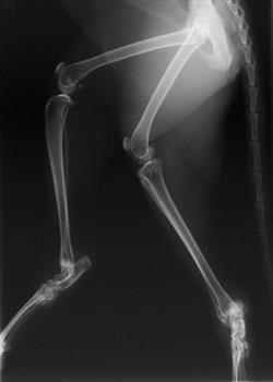
|
|
| |
| | Radiograph 2. | 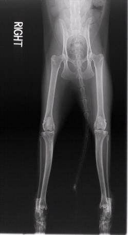
|
|
| |
|
Joint Fluid Examinations
Left tibiotarsal joint aspirate: Smears (2) contain blood and abundant mucoproteinaceous background precipitate typical of synovial fluid. Estimated leucocyte count: 80,000/microliter; leucocytes are neutrophils, the majority of which are nondegenerated; no organisms seen.
Right tibiotarsal joint aspirate: Smears (5) contain blood and abundant mucoproteinaceous background precipitate typical of synovial fluid. Estimated leucocyte count: 60,000/microliter; leucocytes are neutrophils, the majority of which are nondegenerated; no organisms seen.
Right femorotibial joint aspirate: Smears (2) contain blood and abundant mucoproteinaceous background precipitate typical of synovial fluid. Estimated leucocyte count: 2,000-4,000/microliter; leucocytes are neutrophils, the majority of which are nondegenerated; no organisms seen.
[Top]
Case Summary
The signalment, clinical signs, radiographic studies and joint fluid cytology in this case were all consistent with immune-mediated polyarthritis.
The clinical presentation of this case suggested an orthopedic disorder or polyarthritis. The imaging studies did not reveal erosive changes involving the articular joint surfaces. The synovial fluid cytology was also consistent with a polyarthritis. These changes in conjunction with a positive ANA serology suggest that this case fits the category of systemic lupus erythematosus of the immune-mediated polyarthropathies.
There are at least four classes of immune-mediated polyarthropathy described in the cat, including rheumatoid arthritis, progressive proliferative polyarthropathy, systemic lupus erythematosus and idiopathic polyarthritis. Rheumatoid arthritis and progressive proliferative polyarthropathy develop distinguishing radiographic features and can be further classified by the presence or absence of rheumatoid factor. While systemic lupus erythematosus and idiopathic polyarthritis are two nonerosive arthropathies that cause synovial effusion but do not develop any distinguishing radiographic features.
Rheumatoid arthritis and progressive proliferative polyarthropathy are progressive disorders that mainly affect young male cats less than 5 years of age. The carpi and tarsi are the joints mostly likely to be involved, however other joints have been reported with a smaller frequency. The relationship of concurrent viral infections (feline syncytium-forming virus and feline leukemia virus), has been questioned as not all affected cats are positive for feline leukemia virus. These two arthropathies can be further divided into destructive and proliferative forms based on the typical radiographic features that are found.
The last two classes of immune-mediated polyarthropathy, systemic lupus erythematosus and idiopathic polyarthritis, do not produce any distinctive radiographic changes but do present with a symmetric distribution of joint swelling. Systemic lupus erythematosus is distinguished from idiopathic polyarthritis by the presence of a positive antinuclear antibody test. Because of an association between feline idiopathic polyarthritis and myeloproliferative disease, a bone marrow biopsy is recommended for any cat identified with a nonerosive , immune-mediated polyarthritis.
Progressive orthopedic problems in animals can sometimes be very difficult to recognize until they are well advanced. Such cases are sometimes referred to veterinary neurologists because of the vague and often varying nature of the clinical signs.
References
1. Pedersen, NC, Pool, RR, O Brien, T. Feline Chronic Progressive Polyarthritis. J Am Vet Res 1992; Vol 41, No. 4: 522-535.
2. Allan, GS. Radiographic Features of Feline Joint Diseases: in Veterinary Clinics of North America: Small Animal Practice. 2000; Vol 30, No. 2: 281-301
[Top]