 |
|
|
|
An Algorithmic Approach to Superficial Tumor Management
Rodney L. Page MS, DVM; Diplomate ACVIM (Internal Medicine/Oncology)
College of Veterinary Medicine, Cornell University
Clinical Evaluation
General health status is assessed to identify diseases that may adversely affect prognosis, and limit or alter therapy. After a thorough physical examination the screening laboratory evaluation generally includes a complete blood cell count, serum biochemistry panel, and urinalysis. Other diagnostic tests are performed as indicated. Survey radiographs are indicated to detect metastasis, determine potential bone involvement, evaluate orthopedic soundness prior to amputation or limb-sparing in dogs with osteosarcoma, localize oral or nasal masses, etc. Contrast radiographic studies can determine the extent of gastrointestinal and genitourinary neoplasia. Computed axial tomographic (CAT) scanning or magnetic resonance imaging (MR) are becoming more available and defines the invasive characteristics of deep seated tumors much more clearly than survey radiographs. CAT or MR imaging procedures are particularly helpful when planning involved surgical procedures. Ultrasonography can be used to determine the proximity of a tumor to large blood vessels, to determine the cavitary or cystic nature of masses, to evaluate possible intraabdominal metastases to lymph nodes or organs, and to assess the initial and post-treatment tumor volume.
Cytologic examinations of bone marrow aspirates, buffy coat preparations of peripheral blood samples and fine needle aspiration biopsies of accessible tumors and regional lymph nodes are important diagnostic procedures. Fine needle aspiration can be accomplished on any accessible mass. Often, a rapid, inexpensive diagnosis can be made for certain tumor types (lipomas, sebaceous adenomas, mast cell tumors). However, cytologic evaluation of fine needle aspirates or bone marrow specimens must not be over-interpreted. Treatment decisions should be based on a cytologic diagnosis only when a definitive diagnosis can be made such as with lymphosarcoma, mast cell tumors, etc.
Tumor Biopsy
Many techniques are available for tissue biopsy. The method selected should safely and simply procure adequate tissue samples to provide an accurate diagnosis without compromising treatment. Biopsies can be excisional (complete removal of the tumor) or nonexcisional (removal of only a portion of the tumor). Nonexcisional techniques include: a) cytology from a fine-needle aspirate, brush samples, impression smears or effusions, b) histopathology of cutting forcep biopsies, cutting needle biopsies, punch biopsies, and incisional biopsies.
a. Principles of Tumor Biopsy:
In general, excisional biopsy is preferable if the mass is small (<3 cm in diameter), freely moveable, and without adjacent tissue invasion. The specimen must contain a complete margin of normal tissue (preferably 2-3 cm in all directions). Specific indications for excisional biopsy include lymph nodes, small cutaneous nodules with ample surrounding normal tissue, mammary gland tumors, tumors of the central nervous system (to provide decompression) and masses found during a laparotomy or thoracotomy since a re-excision is unlikely.
Incisional biopsy is recommended if a definitive diagnosis or histologic grade would influence the treatment decision. For example, histologic grade of soft tissue sarcomas and mast cell tumors are prognostic factors that can be helpful in treatment planning. Biopsy results may suggest the degree of surgical resection necessary for definitive control or indicate that additional types of therapy may be beneficial.
Ideal histologic samples contain the neoplastic tissue and some adjacent normal tissue to evaluate the tumor boundary. Superficial tumors should be sampled at the tumor/normal tissue interface away from regions of ulceration, necrosis or inflammation. Deep biopsies (> 1 cm) may be necessary to avoid sampling only overlying tissue. Biopsy at the tumor margin can be undesirable in certain deep seated tumors if it disrupts and thereby extends the tumor margin. This can necessitate wider resection or a larger radiation field for adequate treatment. Biopsy needles are preferred for deep seated tumors. The biopsy incision or needle tract for every biopsy should be preplanned since it is a potentially contaminated region, and should be removed at the time of the definitive procedure or included in the radiation treatment field.
A histologic grade can be assigned by a pathologist to each tumor specimen. A histologic grade represents the degree of malignancy and is based on tissue differentiation, mitotic activity and extent of necrosis within the tumor. The grade is often of prognostic significance and can be used to suggest treatment alternatives. If the histologic diagnosis is questionable or does not seem to match your clinical judgement, discuss the results with the pathologist.
Tumor Staging
Staging is used to determine the extent of neoplastic disease, provide a framework for rational treatment planning, facilitate communication between clinicians, allow for uniform comparison and evaluation of treatment results, and aid prognostication. Accurate staging requires an understanding of the biologic behavior of different tumor types and a thorough diagnostic evaluation. Lymph node evaluation is extremely important! Surgical resection of lymph nodes that drain the region have to be incorporated into biopsy and staging evaluations more rigorously than they have in the past.
Several staging systems are available. Most are based on assessment of local, regional, and distant disease involvement. Some systems include other factors such as presence or absence of clinical signs (e.g. lymphomas), histologic grade (e.g. mast cell tumors), or site (e.g. squamous cell carcinoma of mouth, tonsil, pinna, digit). The TNM (Tumor-Node-Metastasis) system devised by the World Health Organization is the standard system for most tumors in veterinary medicine.
General Therapeutic Considerations
Tumor Biology and Natural History
Rational treatment planning involves basic knowledge of the potential for local recurrence and metastasis of the neoplasm. The keystone of this information is the histological assessment or diagnosis. Such information is available in many formats currently (text, WWW). Malignant tumors predisposed to local recurrence should be managed aggressively from the time of initial diagnosis. The chance for long-term tumor control is greatest when the tumor is undisturbed by previous therapeutic intervention. The risks and benefits of aggressive management must be carefully explained. However, most owners will recognize the obvious benefit of prolonged tumor response with reduced overall expense if the tumor can be managed once, albeit initially more costly, compared to multiple, suboptimal attempts at tumor control.
Goals of Treatment
Maintaining the highest quality of life for the longest period of time is always the goal of cancer management in companion animals. This goal must be considered within the context of client emotional and financial restrictions. Decisions are often difficult. The best service that can be provided to a client with a pet that has cancer is a knowledgeable, unbiased assessment of the condition and a frank discussion of options sufficient to allow the client to make an informed decision. This may involve consultation or referral to a specialist or a comprehensive cancer center.
A working definition of 'curative' therapy often used in veterinary medicine is the likelihood of >50 % that a given tumor type will be controlled for at least 1 year following treatment. If the best available information suggests this is not possible, palliative therapy may be considered.
The first therapeutic determination made by the veterinarian involves whether the tumor may be completely excised. This is determined based on the size of the surgical field necessary to remove all known and probable tumor extent, the site of the tumor and the skill of the surgeon. The site of the tumor dictates the extent of normal tissue resection. For instance, interscapular injection-site sarcomas in cats require extensive removal of tissue, including portions of dorsal vertebral processes and scapulae, due to the complex nature of the fascial planes within that site. Regions where sufficient normal tissue cannot be removed (e.g., distal extremities, skull) may require extensive reconstruction (grafting) or consideration of multimodality therapy. More sophisticated tumor imaging techniques, such as CT or MR, greatly assist presurigcal planning for invasive tumors or for tumors located close to critical normal structures. The skill and experience of the surgeon is extremely important. Removal of tumors and reconstruction of normal tissue in difficult locations requires skills that must be continuously practiced and updated. Some tumors are radiation sensitive (e.g., acanthomatous epulis, plasma cell tumors, mast cell tumors) and may be considered potentially curable if they are located within a site that is not amenable to complete resection. A combination of radiation and surgery improves outcome in situations when neither treatment modality alone is sufficient to accomplish that goal. Well planned, combined modality therapy is being used more frequently for tumors which are located in difficult sites.
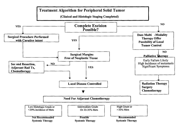
Figure 1 illustrates the process of determining the treatment options for a solid tumor.
|
The pivotal point in this algorithm is the assessment of the normal tissue margins surrounding the tumor. This is critical information that is used to define the prognosis and treatment options. Assuming the surgery was attempted because it was deemed feasible to resect the mass in its entirety by the removal of generous normal tissue boundaries the issue becomes a microscopic determination of contamination. The pathologic evaluation of the specimen will consist of several sections obtained across the entire tumor. Extensive sectioning is not done routinely therefore, identifying the areas of greatest concern for the pathologist to focus the microscopic evaluation on is prudent. Tissue marking dyes are readily available and inexpensive. On-line instructions and templates are available at www.bradleyproducts.com.
The need for adjuvant therapy is based on a high likelihood of local tumor recurrence following resection or a high rate of metastasis even if the primary tumor is permanently controlled. Adjuvant radiation therapy is recommended for local control of incompletely resected sarcomas or mast cell tumors and results in long term control. Adjuvant chemotherapy or adjuvant immunotherapy would be theoretically valuable for any tumor with a substantial metastatic rate. Tumors that are associated with an incidence of distant metastasis exceeding 20% may warrant a recommendation for adjuvant treatment if a survival benefit could be documented for that chemotherapeutic protocol. In veterinary medicine, few studies have documented that chemotherapy is associated with prolonged survival in the adjuvant setting. Survival of dogs with osteosarcoma and, perhaps, hemangiosarcoma, is significantly prolonged after chemotherapy. Cats with mammary carcinoma are believed to benefit from adjuvant chemotherapy. A general recommendation for adjuvant therapy in other types of cancer where metastasis is a life-limiting event is difficult to make given the available data. However, some types of cancer (high-grade sarcoma) are associated with a high risk of metastasis and clinical studies are being conducted to determine efficacy of adjuvant therapy in these tumor categories.
|
|

Copyright ACVC

